A Doctor’s View: What Happens in a Glaucoma Exam
Posted in Bellingham, Eye Blog, Eye Exam, Fluctuating Vision, Medical, Mount Vernon, OCT test, Whidbey
By Ernesto Golez MD
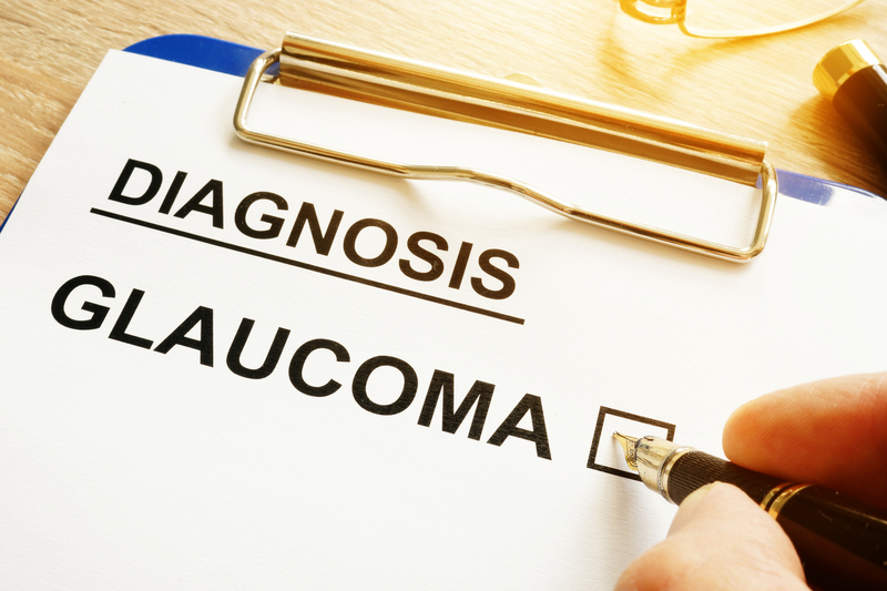
It can be disquieting to find out you have glaucoma, but seeing a specialist for a glaucoma exam regularly can help slow the progression of symptoms. As a glaucoma specialist, I see many patients who are curious to learn what is different about glaucoma eye care and what happens when a they come in to have a glaucoma evaluation.
A medical glaucoma exam includes a few steps:
Taking Histories
For a new patient, a more comprehensive evaluation may be needed. In the examination room, you will be asked about current eye complaints related to glaucoma, including eye pain, loss of peripheral and central vision, blurry vision, headaches, and halos.
Your doctor will ask several follow-up questions, including when the symptoms started and how long and how severe they are. They will also ask about past medical history, including diagnoses like diabetes, hypertension, hypotension, sleep apnea, migraine, and history of steroid use. They will also discuss family medical history, especially of glaucoma.
At a glaucoma exam you may also have your blood pressure and pulse checked.
Refraction for Visual Acuity
Your current refractive error is often obtained (checking your vision with or without glasses). This will eliminate other diagnoses of blurry vision due to refractive state, as well as to help determine your glaucoma risk factors. For example, far-sighted people (those who need glasses for reading before age 40) may have smaller eyeballs that can cause angle-closure glaucoma. Near-sighted people (those who need glasses for distance) may have optic nerves that falsely signal glaucoma, so having their visual acuity test helps determine this.
External Examination and Pressure Check
The external examination of the eye will be next. This includes tests for pupil reaction, peripheral vision testing, eye movement and overall appearance of the eye. Eye pressures will also be taken, since elevated eye pressure is one of the major risk factors of glaucoma. Another important diagnostic is determining the thickness of the cornea (the clear outer area of the eyeball). Cornea thickness may affect how pressures are read. Thick corneas may create a falsely high eye pressure, while thin corneas may create a falsely low eye pressure. This is measured using a pachymeter.
Slit Lamp Exam
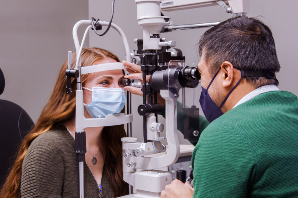
The doctor also uses an instrument called a slit lamp biomicroscope to examine the eye in magnified view. While on the slit lamp, the doctor will be using special lenses to look at the insides of the eye. One of the lenses is called the gonioscope, which views the anterior chamber angle to determine if the fluid drain of the internal eye is open or closed. Another lens is used to evaluate the optic nerve and other structures such as the macula and the retina.
Patients may need to be dilated to see the internal structures more clearly, especially the peripheral retina.
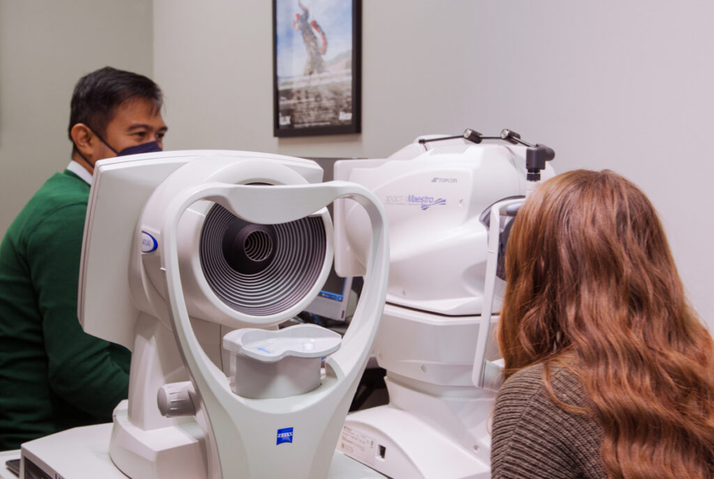
Optic Nerve Check
Documentation of the optic nerve is also helpful to determine structural damage and progressive damage of the nerve. Examples of diagnostic tests that help document appearance of the optic nerve are a stereo disc photograph, and OCT (ocular coherence tomography) and an HRT (Heidelberg Retinal Tomography). These newer devices can detect structural damage early so treatment can be started.
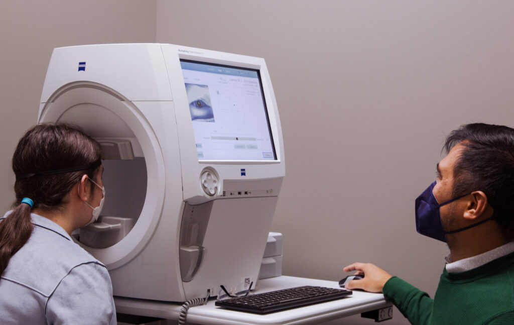
Visual Field Test
Since glaucoma causes permanent vision loss, it is also important to evaluate the patient’s peripheral vision. This is done by using a visual field. The device uses different light intensities and light point sizes at various points in the retina (static perimetry). Light points move from the edge of the patient’s eye towards the center of their vision field until the patient indicates they can see it (kinetic perimetry).
Both optic nerve and visual field testing are performed periodically to determine if glaucoma is progressing. It also tells whether a treatment is working or not. Frequency of testing will depend on the severity of the disease and accuracy of the test.
Click here to learn more about testing.
Follow Up Appointments
At the end of each glaucoma exam, the physician will review his diagnosis, go over a treatment plan, and prescribe the timing for any follow-up appointments.
Contact Cascadia Eye
If you would like to learn more about glaucoma, or if you would like to schedule a consultation at Cascadia Eye, please contact us today. We are happy to answer any questions you might have!
In addition, join us on Facebook, Instagram or YouTube to ask your questions about eyes, exams, and our practice. We’d love to hear from you – and there might be a blog to address your questions in the future.